


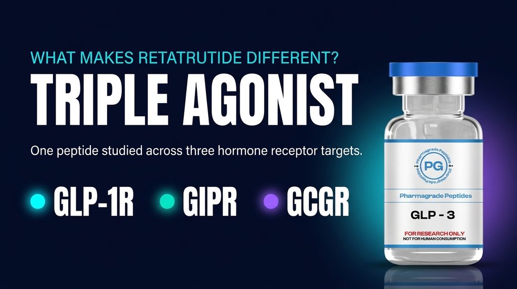
Retatrutide is a research peptide studied for its ability to activate three different hormone receptors: the glucose-dependent insulinotropic polypeptide receptor (GIPR), glucagon-like peptide-1 receptor (GLP-1R), and glucagon receptor (GCGR). This three-receptor activity makes its molecular design different from compounds that act on only one or two of these targets.
Understanding Retatrutide Receptor Selectivity requires looking beyond the phrase triple agonist. At PharmaGrade Peptides, we explore the scientific research behind how retatrutide interacts with each receptor, which molecular features support those interactions, and why its activity differs between targets. Structural studies published in Cell Discovery in 2024 provide important details through cryo-electron microscopy and receptor mutation experiments.
This article examines those findings, explains how receptor activity is measured, and identifies the limits of current evidence.

What Does Retatrutide Receptor Selectivity Mean?
Receptor selectivity describes how a molecule interacts with different biological receptors. In peptide research, scientists examine whether a compound binds to a receptor, activates it, and produces a measurable cellular response.
Retatrutide is described as a triple agonist because it activates three receptors:
- GLP-1R: Glucagon-like peptide-1 receptor
- GIPR: Glucose-dependent insulinotropic polypeptide receptor
- GCGR: Glucagon receptor
These receptors belong to the class B1 G protein-coupled receptor (GPCR) family. They respond to different peptide hormones and participate in several biological signalling pathways.
However, triple agonism does not mean that retatrutide interacts with all three receptors equally. Each receptor has a different molecular structure, and the peptide can form different interactions within each receptor’s binding pocket.
Therefore, Retatrutide Receptor Selectivity involves understanding both its shared activity across the three targets and the differences in its interactions with each one.
The Three Receptors Targeted by Retatrutide
Retatrutide’s molecular profile includes three distinct receptor targets. Each receptor has its own structure and responds to specific peptide signals.
1. GLP-1 Receptor (GLP-1R)
The GLP-1 receptor belongs to the class B GPCR family. It responds naturally to glucagon-like peptide-1, a hormone involved in glucose regulation and other physiological processes.
In molecular research, scientists examine how peptide agonists interact with the receptor’s extracellular region and transmembrane binding pocket. These interactions help explain how a peptide can stabilise receptor conformations associated with cellular signalling.
Retatrutide activates GLP-1R, but its interaction is not identical to that of native GLP-1 or other GLP-1 receptor agonists. Its amino-acid sequence and structural features allow it to interact with multiple receptor types.
2. GIP Receptor (GIPR)
The GIP receptor responds to glucose-dependent insulinotropic polypeptide. Like GLP-1R, it is a class B GPCR and participates in hormone-mediated signalling.
GIPR is an important part of retatrutide’s three-target profile. Structural research shows that retatrutide can form interactions with receptor-specific amino-acid residues while maintaining some interactions shared across the three target receptors.
The receptor’s extracellular loops also contribute to peptide recognition. Differences in these regions help explain why the same peptide can interact with GIPR differently from GLP-1R or GCGR.
3. Glucagon Receptor (GCGR)
The glucagon receptor responds naturally to glucagon and belongs to the same GPCR family.
Its inclusion gives retatrutide a third receptor target beyond GLP-1R and GIPR. However, GCGR has structural differences that affect how peptide molecules interact with its binding pocket.
The 2024 structural study identified receptor-specific contacts between retatrutide and GCGR. These findings help researchers understand how a single peptide sequence can accommodate the molecular features of three different receptors.
Does Retatrutide Activate All Three Receptors Equally?
No. Retatrutide activates all three receptors, but its measured activity differs between them.
This distinction is important when discussing Retatrutide Receptor Selectivity. The term triple agonist describes the number of receptor targets, not an equal level of activity at each target.
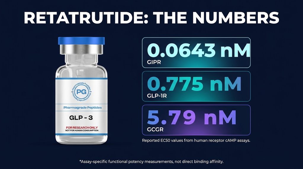
A 2026 review reports the following half-maximal effective concentration (EC50) values from human receptor cAMP assays:
| Receptor | Reported EC50 |
| GIPR | 0.0643 nM |
| GLP-1R | 0.775 nM |
| GCGR | 5.79 nM |
In these specific assays, the reported EC50 was lowest at GIPR and highest at GCGR. A lower EC50 indicates that a lower concentration was required to produce half of the measured maximum response under the conditions of that experiment.
These values should not be interpreted as universal measures of receptor preference. They are assay-dependent functional potency measurements. They do not, by themselves, establish receptor occupancy, tissue-level activity, or the strength of every possible signalling pathway.
The experimental conditions also matter. Receptor expression, cell type, assay design and other factors can influence measured potency. Researchers therefore need more than one type of experiment to build a complete picture of receptor activity.
How Structural Research Explains Retatrutide’s Three-Target Activity
One of the most important studies on Retatrutide Receptor Selectivity was published by Li and colleagues in Cell Discovery in July 2024.
The researchers used cryo-electron microscopy (cryo-EM) to examine retatrutide in complex with GLP-1R, GIPR and GCGR. They also investigated receptor mutations to understand which amino-acid residues contributed to peptide activity.
The study revealed that retatrutide uses a combination of shared and receptor-specific molecular interactions.

Conserved interactions across the three receptors
Some amino-acid residues within the three receptors have similar structural positions and chemical properties. Retatrutide can interact with these conserved regions through different types of molecular contacts.
The study identified interactions that included:
- Hydrogen bonds
- Salt bridges
- Hydrophobic contacts
- Aromatic stacking interactions
These interactions help explain how one peptide sequence can be recognised by three related receptors.
However, the interactions are not identical in every receptor. Small differences in amino-acid residues and their positions can change how the peptide fits within each binding pocket.
Receptor-specific interactions
The study also identified differences in the extracellular loops and other receptor regions.
Extracellular loops are flexible parts of GPCR structures that help shape the receptor’s binding environment. Their positions and conformations can vary between receptor types.
For retatrutide, these differences help explain how the peptide accommodates GLP-1R, GIPR and GCGR despite their structural similarities.
The researchers also used mutagenesis experiments. In these experiments, selected receptor amino acids were changed to examine how those changes affected retatrutide-induced signalling.
When certain residues were altered, the measured cAMP response changed. This provided additional evidence that individual molecular contacts contribute to receptor recognition and activity.

Why Amino-Acid Sequence Matters in Receptor Recognition
Peptides are made from amino acids arranged in a specific sequence. That sequence influences molecular shape, chemical properties and the way a peptide interacts with its target.
For a multi-receptor peptide such as retatrutide, sequence design is particularly important. The molecule must accommodate the structural features of more than one receptor.
The 2024 cryo-EM research found that retatrutide adopts a continuous helical structure when bound to the three receptors. Its N-terminal region enters the receptor’s transmembrane domain, while other parts of the peptide interact with extracellular receptor regions.
The study also highlighted differences in the way the middle and C-terminal regions interact with receptor-specific structures.
This helps explain why peptide sequence design is not simply about adding more receptor targets. Small changes in amino-acid composition can influence molecular recognition, functional potency and receptor signalling.
For researchers studying peptide structure, these findings provide a framework for examining how sequence modifications affect interactions with related GPCRs.
How Researchers Measure Retatrutide Receptor Activity
Scientists use several experimental methods to investigate receptor interactions. No single method provides every detail about binding, activation and signalling.
1. Cryo-electron microscopy
Cryo-EM allows researchers to examine the structures of biological molecules at high resolution. Samples are rapidly frozen, and images are collected to reconstruct three-dimensional molecular structures.
In retatrutide research, cryo-EM helped reveal how the peptide sits within the binding environments of GLP-1R, GIPR and GCGR.
The resulting structures provide information about molecular positioning and potential interactions between peptide and receptor residues.
2. cAMP signalling assays
Cyclic adenosine monophosphate (cAMP) is an intracellular signalling molecule. Class B GPCR activation can stimulate pathways that change cellular cAMP levels.
Researchers use cAMP assays to measure receptor activation after exposing receptor-expressing cells to a test compound.
These experiments can help estimate functional potency through EC50 values. However, cAMP is only one signalling readout. It does not describe every possible receptor response.
3. Receptor mutagenesis
Mutagenesis involves changing selected amino acids within a receptor. Researchers then compare the response of the modified receptor with that of the original receptor.
If a mutation changes the measured response to retatrutide, the affected residue may contribute to receptor recognition or activation.
This method helped the 2024 research team investigate the importance of specific molecular contacts across the three receptors.
4. Radioligand binding assays
Binding assays examine how a molecule interacts with a receptor. Depending on the experimental design, they can help estimate binding affinity or compare how different compounds engage a target.
Binding affinity and functional potency are related concepts, but they are not interchangeable. A compound may bind to a receptor without producing the same level of functional activation as another compound.
Using structural studies, binding experiments and functional assays together provides a more complete understanding than relying on one measurement alone.
Receptor Binding vs Functional Potency: What Is the Difference?
These terms are sometimes used as if they mean the same thing, but they describe different experimental properties.
Receptor binding refers to the interaction between a molecule and a receptor. Binding studies may estimate how strongly a compound associates with its target.
Functional potency describes the concentration required to produce a defined biological response in a particular assay. EC50 is a commonly reported measure of functional potency.
A lower EC50 does not automatically mean stronger binding affinity. The measured response can also depend on receptor expression, signalling amplification, cell type and assay conditions.
This distinction matters when interpreting Retatrutide Receptor Selectivity. Comparing EC50 values across different receptor systems requires care, particularly when the assays use different experimental conditions.
A reliable interpretation should identify the assay, receptor system, measured response and experimental limitations.
What Are the Limitations of Current Receptor Selectivity Research?
Structural and functional studies have improved scientific understanding of retatrutide, but several limitations remain.
Laboratory findings do not explain every biological response
Cryo-EM structures show molecular arrangements under specific experimental conditions. They do not reproduce every feature of a living biological system.
Cell-based assays also have limitations. Receptor expression levels and cellular signalling machinery can affect experimental results.
Receptor activity can vary between species
A peptide may interact differently with human and animal receptors. Researchers should not assume that results from one species apply directly to another.
This is particularly important when interpreting preclinical research.
One signalling pathway cannot represent every receptor response
cAMP assays provide useful information about G protein signalling. However, GPCRs can activate multiple signalling pathways.
Other measurements may be needed to examine beta-arrestin recruitment, receptor internalisation, desensitisation and additional cellular responses.
Structural similarity does not mean identical function
GLP-1R, GIPR and GCGR belong to the same receptor family, but they have different molecular features. Similar overall structures do not mean that every peptide interaction produces the same functional result.
More research is needed to understand how molecular interactions relate to broader biological responses.
Frequently Asked Questions About Retatrutide Receptor Selectivity
What receptors does retatrutide target?
Retatrutide is a triple agonist studied for activity at three receptors: GLP-1R, GIPR and GCGR. These receptors belong to the class B1 GPCR family.
Is retatrutide selective for one receptor?
Retatrutide activates all three target receptors, but its measured functional potency differs between them. The results depend on the assay and experimental conditions, so it is more accurate to describe its activity using specific receptor and assay data rather than a single universal selectivity label.
Which receptor shows the lowest reported EC50 for retatrutide?
A 2026 review reports the lowest EC50 value at GIPR in the cited human receptor cAMP assays. The reported values were 0.0643 nM at GIPR, 0.775 nM at GLP-1R and 5.79 nM at GCGR. These are assay-specific functional potency measurements, not universal measures of receptor binding.
How did researchers study retatrutide’s receptor interactions?
A 2024 Cell Discovery study used cryo-electron microscopy to examine retatrutide bound to GLP-1R, GIPR and GCGR. The researchers also used receptor mutagenesis and cAMP signalling experiments to investigate important molecular interactions.
What is the difference between a dual agonist and a triple agonist?
A dual agonist activates two receptor targets, while a triple agonist activates three. Retatrutide is studied as a triple agonist because it activates GLP-1R, GIPR and GCGR.
Does an EC50 value measure receptor binding affinity?
No. EC50 measures functional potency in a particular assay. Binding affinity is assessed through separate binding experiments. The two measurements provide different information and should not be treated as interchangeable.
Why is cryo-EM important in retatrutide research?
Cryo-EM helps researchers examine the three-dimensional structure of peptide-receptor complexes. It can reveal how a peptide is positioned within a receptor and identify molecular regions that may contribute to recognition and activation.
Conclusion
Retatrutide Receptor Selectivity is best understood through the relationship between its peptide sequence, receptor structure, and measured functional activity.
Research identifies GLP-1R, GIPR, and GCGR as its three receptor targets. The 2024 cryo-EM study showed that retatrutide can form shared molecular contacts across these receptors while also accommodating receptor-specific structural differences. Functional assays provide another layer of evidence, showing that measured potency is not identical across the three targets.
These findings make retatrutide an important subject in multi-receptor peptide research. However, structural observations and laboratory measurements have defined limits. Binding affinity, functional potency and broader biological outcomes must be evaluated separately.
Researchers can buy Retatrutide Glp-3 from PharmaGrade Peptides for better research results and laboratory research purposes.
Research-use notice: This article is provided for scientific and educational discussion. It does not provide medical advice or instructions for human or veterinary use. Experimental findings should be interpreted within the limits of the methods and evidence reported in the cited research.
References
- Li, W. et al. (2024). “Structural insights into the triple agonism at GLP-1R, GIPR and GCGR manifested by retatrutide.” Cell Discovery, 10, Article 77. Read the research paper.
- Li, W. et al. (2024). PubMed record for the structural study. View publication details.
- “Retatrutide as a Unimolecular GIP/GLP-1/Glucagon Receptor Tri-Agonist: Molecular Design, Pharmaceutical Development and Critical Interpretation of Emerging Phase 3 Evidence.” Pharmaceutics (2026). Read the review.
- “Progress in Anti-Obesity Drug Development: A Pharmacodynamics Perspective” (2026). Read the review.
